The Parasympathetic Nervous System: Structure, Function, and Physiological Role

Medical illustration of the parasympathetic nervous system showing craniosacral outflow pathways extending from the brainstem and sacral spinal cord to internal organs.

Table of Contents

The parasympathetic nervous system (PNS) represents one of the two primary divisions of the autonomic nervous system (ANS), the intricate network responsible for regulating the body’s involuntary functions. Working in concert with its counterpart, the sympathetic nervous system (SNS), the PNS is instrumental in maintaining the body’s internal balance, a state known as homeostasis
 
While it is often described by the simplified moniker “rest and digest,” this phrase only captures a fraction of its profound and continuous physiological influence. A more precise understanding reveals the PNS as a critical modulator of energy conservation, recovery, and the baseline operations essential for life.
 
This article provides a comprehensive, university-level overview of the parasympathetic nervous system, adapted for an educated general readership. It explores the system’s anatomical structure, its intricate signaling mechanisms, its core physiological functions, and its role in stress recovery and interaction with other neural systems. 
 
The content is grounded in established neurobiological and physiological research, avoiding medical advice or experiential language to maintain a neutral, academic, and evidence-based perspective.
 

What Is the Parasympathetic Nervous System?

 
The parasympathetic nervous system is a division of the autonomic nervous system that primarily directs the body’s resources toward functions of conservation and restoration. It counterbalances the sympathetic nervous system, which orchestrates the “fight or flight” response to stressful or high-exertion situations. 
 
In contrast, the PNS governs activities that allow the body to rest, recover, and process nutrients, thereby ensuring long-term physiological stability. Its actions are generally aimed at slowing down high-energy functions and promoting processes that occur when the body is at ease.
 
This system is fundamental to homeostasis, the dynamic equilibrium of the body’s internal environment. For instance, after a period of stress where the heart rate is elevated, the PNS actively works to decrease it, returning the cardiovascular system to a baseline state. The popular term “rest and digest” refers to its role in decreasing heart rate, lowering blood pressure, and stimulating digestive processes, including salivation, gut motility, and the release of digestive enzymes
 
However, its functions extend far beyond digestion, encompassing pupillary constriction, bladder control, and sexual arousal, among other vital processes. The balance between the parasympathetic and sympathetic systems is not merely oppositional but a highly integrated and dynamic interplay that allows the organism to adapt to a constantly changing internal and external environment.
 

The Autonomic Nervous System: Context and Organization

The autonomic nervous system operates largely below the level of conscious control, managing the essential machinery of the body. It is broadly divided into the sympathetic and parasympathetic divisions, which together regulate the function of visceral organs. While the sympathetic nervous system prepares the body for action by mobilizing energy reserves, the parasympathetic nervous system acts to conserve energy and oversee routine operations. 
 
This is not a simple on-off switch; rather, the two systems provide tonic, continuous input to target organs, with their relative influence shifting based on physiological demands.
 
The central control of the ANS originates in higher brain centers, most notably the hypothalamus and various nuclei within the brainstem. These regions process sensory information from the body and the external environment to orchestrate appropriate autonomic responses. The commands are then relayed through two-neuron pathways to the target organs. This pathway consists of a preganglionic neuron, whose cell body is in the central nervous system (CNS), and a postganglionic neuron, whose cell body is in a peripheral ganglion.
 
The parasympathetic division is characterized by its craniosacral outflow, meaning its preganglionic neurons originate in specific cranial nerve nuclei in the brainstem and in the sacral region of the spinal cord (segments S2–S4) . This anatomical arrangement is distinct from the thoracolumbar outflow of the sympathetic system. The balance between these systems is a model of complex integration, ensuring that organ function is finely tuned to meet the body’s needs at any given moment.
 

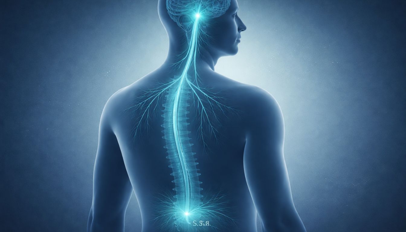
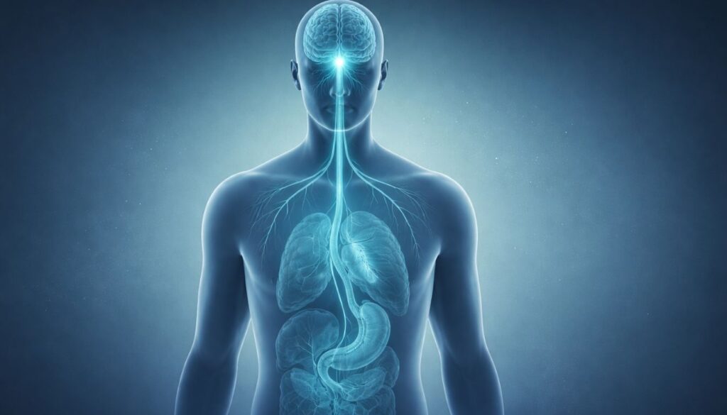
Anatomical Pathways of the Parasympathetic System


Medical-style illustration of the vagus nerve extending from the brainstem to the heart and digestive organs, representing parasympathetic control.
The vagus nerve carries the majority of parasympathetic fibers, connecting the brainstem to the heart, lungs, and digestive tract to regulate rest-and-digest functions.
The anatomical organization of the parasympathetic nervous system is defined by the origin of its preganglionic neurons and the length of its neural fibers. The system’s preganglionic neurons have their cell bodies located in two distinct regions of the central nervous system: the brainstem and the sacral spinal cord. 
 
The cranial portion of the PNS outflow involves four cranial nerves: the Oculomotor Nerve (III), which controls pupillary constriction; the Facial Nerve (VII), which governs lacrimal and salivary glands; the Glossopharyngeal Nerve (IX), which innervates the parotid salivary gland; and the Vagus Nerve (X). The vagus nerve is the most extensive of these, carrying approximately 75% of all parasympathetic fibers and innervating nearly all thoracic and abdominal viscera .
 
The sacral portion of the PNS outflow originates from neurons in the S2, S3, and S4 segments of the spinal cord. These preganglionic fibers form the pelvic splanchnic nerves, which innervate the pelvic viscera, including the lower colon, rectum, bladder, and reproductive organs. 
 
A defining feature of the PNS is its use of long preganglionic fibers and short postganglionic fibers. The preganglionic axons travel from the CNS almost all the way to the target organ before synapsing in a ganglion located on or very near the organ wall. 
 
Consequently, the postganglionic fibers are very short, relaying the signal over a minimal distance. This arrangement allows for highly specific and localized control over target organ function, in contrast to the more diffuse activation often seen in the sympathetic system .
 

Neurotransmitters and Signaling Mechanisms

 
The signaling within the parasympathetic nervous system is primarily mediated by the neurotransmitter acetylcholine (ACh). Unlike the sympathetic system, which predominantly uses norepinephrine at its target organs, the PNS uses ACh at both its preganglionic and postganglionic synapses. 
 
When a preganglionic neuron is activated, it releases ACh that binds to nicotinic receptors on the postganglionic neuron, causing it to fire. The postganglionic neuron then releases ACh at the target organ, where it binds to muscarinic receptors on the effector cells.
 
Muscarinic receptors, which are G-protein coupled receptors, come in five main subtypes (M1-M5). Their activation triggers varied cellular responses depending on the receptor subtype and the target tissue. For example, M1, M3, and M5 receptors typically produce excitatory effects like smooth muscle contraction, while M2 and M4 receptors are often inhibitory, as seen in the heart where M2 receptor activation slows the heart rate
 
This receptor diversity allows ACh to produce a wide range of precise effects. The action of acetylcholine is terminated rapidly by the enzyme acetylcholinesterase, which ensures that the signal is brief and allows for fine-tuned control over visceral functions .
 

Core Physiological Functions

 
The parasympathetic nervous system orchestrates a wide array of physiological processes essential for maintaining the body’s baseline functions and conserving energy. Its influence is widespread, touching nearly every major organ system.
 

Cardiovascular Regulation

One of the most critical roles of the PNS is the regulation of cardiovascular activity. Through the vagus nerve, the PNS exerts a tonic, inhibitory influence on the heart, a phenomenon known as vagal tone. When M2 muscarinic receptors in the heart’s sinoatrial (SA) node are stimulated by acetylcholine, the rate of pacemaker firing decreases, resulting in a lower resting heart rate
 
Furthermore, the PNS slows the conduction of electrical impulses through the atrioventricular (AV) node, contributing to a controlled and regular heart rhythm. High vagal tone is often considered a marker of cardiovascular health and efficient autonomic regulation .
 

Respiratory Effects

 
In the respiratory system, parasympathetic activation leads to bronchoconstriction, or the narrowing of the airways, through the stimulation of M3 receptors. This action helps to regulate airflow under resting conditions and protect the airways from irritants. Additionally, the PNS stimulates the secretion of mucus in the bronchial tree, which is essential for trapping foreign particles and keeping the airway surfaces moist. This function is a normal part of the body’s homeostatic regulation.
 

Digestive Processes

 
The parasympathetic nervous system is the primary driver of digestive activity. It increases gut motility by stimulating peristalsis and promotes the relaxation of digestive sphincters. Furthermore, the PNS enhances digestive secretions, including salivation, gastric acid, and enzymes from the stomach and pancreas. It also promotes the contraction of the gallbladder to release bile. This coordinated effort ensures the efficient processing and absorption of nutrients.
 

Pupillary Constriction

 
The PNS also controls the size of the pupil. Via the oculomotor pathways of cranial nerve III, it stimulates the sphincter muscle of the iris, causing the pupil to constrict (miosis). This reflex is important for protecting the retina from bright light and for sharpening focus on near objects, demonstrating the precise control exerted by the PNS.
 

The Vagus Nerve and Parasympathetic Regulation

 
Central to the function of the parasympathetic nervous system is the vagus nerve. As the longest and most complex of the cranial nerves, it is the primary conduit for parasympathetic signals to the visceral organs, making it a critical component of the brain-gut axis. This bidirectional communication pathway allows the central nervous system to monitor and regulate visceral function. 
 
The influence of the vagus nerve on the heart is particularly significant and is often quantified by measuring heart rate variability (HRV). A higher HRV is generally indicative of greater vagal tone and a well-functioning parasympathetic regulatory system, which has been linked to better stress resilience and overall health .
 

Parasympathetic Nervous System and Stress Recovery

 
While the sympathetic nervous system initiates the acute stress response, the parasympathetic nervous system is essential for the recovery phase that follows. Once a perceived threat has passed, the PNS actively works to counteract the physiological arousal produced by the SNS.
 
This process involves the downregulation of sympathetic activity and the physiological restoration of the body to a state of calm. A healthy and responsive PNS allows for a rapid and efficient return to baseline after a stressful event, preventing the prolonged physiological activation that can lead to chronic stress and its associated health problems
udible banner featuring neuroscience and brain research audiobooks including The Brain That Changes Itself, Why We Sleep, Behave, Incognito, and Livewired.
Curated neuroscience audiobooks from leading researchers and institutions, available through Audible.

Interaction With the Enteric Nervous System

 
The parasympathetic nervous system maintains a crucial relationship with the enteric nervous system (ENS), the intricate network of neurons embedded within the walls of the gastrointestinal tract. The PNS, primarily through the vagus nerve, provides the dominant excitatory input to the ENS, stimulating gut motility, secretion, and blood flow. 
 
Preganglionic parasympathetic fibers synapse on neurons within the enteric plexuses, which then carry out the specific functions of regulating peristalsis and secretion . This hierarchical control is a cornerstone of the brain-gut axis, and neurotransmitters like serotonin, which is abundant in the gut, play a significant role in this communication.
 

Parasympathetic Activity in Research Contexts

 
The study of the parasympathetic nervous system has advanced significantly with the use of non-invasive measurement techniques like heart rate variability (HRV). The high-frequency component of HRV is widely accepted as an index of cardiac vagal tone . Researchers use HRV to investigate autonomic balance and its relationship with various physiological and psychological states. 
 
Reduced HRV is associated with numerous chronic conditions, suggesting a state of autonomic dysregulation. Vagal tone research and neuroimaging findings continue to map the central neural networks that control autonomic outflow, providing a more complete picture of how the brain regulates visceral function.
 

Misconceptions and Oversimplifications

The parasympathetic nervous system is often subject to oversimplifications. Labeling it purely as the “relaxation system” is inaccurate, as it is continuously active in regulating baseline physiological functions. Another misconception is that the parasympathetic and sympathetic systems function in simple opposition. 
 
Their relationship is better described as a complex integration model, where both systems are often simultaneously active and their interplay is finely modulated to produce a context-appropriate response.
 

Current Research Questions

 
Contemporary research on the parasympathetic nervous system is exploring several key areas. One major focus is on individual variability in parasympathetic function and its relation to health and disease. 
 
Another critical area of inquiry is autonomic dysregulation and its role in chronic illness. The field of neurocardiology is specifically examining the intricate neural communication between the heart and the brain, while psychophysiology research continues to explore the links between parasympathetic activity, emotion, and cognition.
1️⃣ What is the parasympathetic nervous system responsible for?

The parasympathetic nervous system regulates energy conservation, recovery, and baseline physiological functions. It slows the heart rate, stimulates digestion, promotes glandular secretion, and supports restorative processes essential for homeostasis.

The parasympathetic nervous system conserves energy and supports recovery, while the sympathetic nervous system mobilizes energy during stress or exertion. Rather than functioning as simple opposites, both systems continuously interact to maintain physiological balance.

“Rest and digest” is a simplified phrase describing parasympathetic activation. It refers to decreased heart rate, increased digestive activity, and physiological processes that occur when the body is not under acute stress.

The parasympathetic nervous system has a craniosacral outflow. Its preganglionic neurons originate in brainstem nuclei (cranial nerves III, VII, IX, and X) and in the sacral spinal cord segments S2–S4.

The vagus nerve carries the majority of parasympathetic fibers to thoracic and abdominal organs. It plays a major role in regulating heart rate, digestion, and communication along the brain–gut axis.

The parasympathetic nervous system primarily uses acetylcholine at both preganglionic and postganglionic synapses. It binds to nicotinic receptors in ganglia and muscarinic receptors at target organs.

Parasympathetic activation decreases heart rate by stimulating M2 muscarinic receptors in the sinoatrial (SA) node. This inhibitory influence is often referred to as vagal tone.

Heart rate variability measures fluctuations in time between heartbeats. The high-frequency component of HRV is commonly used as an index of cardiac parasympathetic (vagal) activity in research settings.

No. The parasympathetic nervous system is continuously active, providing tonic regulation of organs. Its influence increases during recovery periods but does not switch off completely during stress.

After a stress response initiated by the sympathetic nervous system, the parasympathetic nervous system helps restore baseline conditions by reducing heart rate, promoting digestion, and stabilizing physiological functions.

Glossary Summary

 
To recap the core concepts covered in this article, the parasympathetic nervous system is a vital component of the autonomic nervous system that governs the body’s “rest and digest” functions. Its key characteristics include:
 
Definition: The division of the ANS responsible for conserving energy and restoring physiological balance.

Anatomy: Characterized by a craniosacral outflow, with long preganglionic and short postganglionic fibers.

Neurotransmitters: Primarily uses acetylcholine (ACh).

Core Functions: Includes slowing the heart rate, aiding digestion, and constricting the pupils.

Research Relevance: Studied extensively through measures like heart rate variability (HRV).
 

Medical and Educational Disclaimer

 
This article is intended for educational and informational purposes only and is not a substitute for professional medical advice, diagnosis, or treatment. Always seek the advice of a qualified health provider with any questions you may have regarding a medical condition.
 

References


Share this post :

Facebook
Twitter
LinkedIn
Pinterest
Popular Product