Brain Regions: Structure, Function, and Scientific Classification

Illustration of major brain regions including frontal, parietal, temporal, and occipital lobes with network connectivity background.

Table of Contents

Introduction

 
The term “brain regions” is a fundamental concept in neuroscience, referring to distinct areas of the brain that are structurally and functionally specialized. Scientifically, a brain region is a defined volume of brain tissue characterized by its unique cellular architecture, connectivity, and physiological role. These regions are not isolated units but are intricately connected, forming complex networks that underpin all aspects of our mental and physical lives. 
 
The classification of brain regions can be approached from both a structural and a functional perspective, and understanding the distinction between these two is crucial for a comprehensive understanding of brain organization. Structural definitions focus on the physical and anatomical characteristics of brain tissue, such as the arrangement of neurons and their connections, while functional definitions are based on the specific roles that different areas play in cognition, emotion, and behavior.
 
In recent years, neuroscience has increasingly shifted towards a network-based perspective of brain function. This approach emphasizes that complex cognitive processes emerge from the coordinated activity of multiple, interconnected brain regions working together as large-scale networks. This article will explore the scientific terminology and classification systems used by neuroscientists to define and study brain regions. 
 
It will delve into the major cortical and subcortical regions, the concept of brain networks, and the role of brain regions in various cognitive functions. Furthermore, it will address the dynamic nature of brain organization through the lens of neuroplasticity and discuss the implications of regional differences in pharmacology. Finally, the article will address common misconceptions and the limitations of region-based models, providing a nuanced and evidence-based overview of this central topic in neuroscience.
 
This article is intended for educational and informational purposes, providing a glossary-style entry into the complex world of brain regions. It aims to clarify the terminology used in neuroscience research and to foster a deeper appreciation for the intricate organization of the human brain. The information presented here is grounded in scientific research and should not be interpreted as medical advice.
 

How Neuroscientists Define Brain Regions

 
Neuroscientists employ a variety of methods to define and classify brain regions, each offering a different level of analysis. These methods range from large-scale anatomical divisions to microscopic cellular distinctions and activity-dependent functional mapping. Historically, the earliest classifications were based on gross anatomy, dividing the brain into major structures visible to the naked eye. 
 
Modern neuroscience, however, integrates anatomical, functional, and connectivity-based approaches to create a more comprehensive and nuanced map of the brain.
 
Anatomical divisions provide the most fundamental framework for understanding brain organization. At the highest level, the brain is divided into the cerebrum, cerebellum, and brainstem . The cerebrum, the largest part of the brain, is split into two hemispheres, and its surface, the cerebral cortex, is famously folded into gyri (ridges) and sulci (grooves). This cortex is traditionally divided into four major lobes: the frontal, parietal, temporal, and occipital lobes, each associated with a broad set of functions. 
 
These anatomical landmarks provide a consistent and universal map for localizing brain structures. Further anatomical classification is based on cytoarchitectonics, the study of the cellular composition of tissues. Brodmann areas, for example, are a classic instance of this approach, where regions are defined by their distinct neuronal organization and layering, a method that often correlates well with functional specialization.
 
Functional specialization offers another crucial layer of classification. A brain region can be defined by the specific cognitive or behavioral function it supports. For instance, Broca’s area in the frontal lobe is defined by its critical role in speech production, a discovery made by observing patients with specific language deficits following damage to this region. 
 
Modern functional neuroimaging techniques, such as functional magnetic resonance imaging (fMRI) and positron emission tomography (PET), have revolutionized this approach. These methods allow researchers to observe which brain regions become active when a person performs a specific task, such as reading, problem-solving, or experiencing emotions. This has led to the mapping of a wide array of functions to specific cortical and subcortical structures, although, as we will see, this mapping is rarely a simple one-to-one relationship.
 
The evolution of brain imaging has also given rise to imaging-based classification. Advanced computational methods can analyze the vast datasets from fMRI and other imaging modalities to parcellate the brain into distinct regions based on patterns of activity and connectivity. For example, regions can be defined by their unique ‘functional connectivity fingerprint,’ which is the pattern of synchronized activity they share with all other brain regions. 
 
This approach has revealed a complex mosaic of functional areas that often do not perfectly align with traditional anatomical boundaries. These methods provide a data-driven way to define brain regions, moving beyond subjective anatomical landmarks and offering a more objective and detailed map of the brain’s functional architecture.
 
Historically, the classification of brain regions has evolved significantly. Early ideas, such as phrenology, attempted to link personality traits to bumps on the skull, a practice now thoroughly discredited. The modern era began with the careful anatomical and clinical work of neurologists like Broca and Wernicke. 
 
Today, the field is moving from a purely region-centric view to a network-based perspective. While the concept of specialized brain regions remains essential, there is a growing understanding that no region works in isolation. 
 
As a leading neuroscience resource from the National Center for Biotechnology Information (NCBI) states, the brain’s functions arise from the coordinated activity of distributed networks of regions . This network perspective, which we will explore later, represents the current frontier in understanding brain organization.
Visualization of interconnected brain regions demonstrating large-scale brain networks and functional connectivity.
Modern neuroscience emphasizes brain networks, where multiple brain regions interact dynamically rather than functioning in isolation.

Major Cortical Brain Regions

 
The cerebral cortex, the outermost layer of the cerebrum, is the most prominent and evolutionarily recent part of the human brain. It is responsible for the highest-level cognitive functions, including thought, language, memory, and consciousness. 
 
The cortex is traditionally divided into four major lobes, each with a constellation of specialized functions. It is important to remember, however, that these lobes are not independent modules but are richly interconnected, and their functions often overlap and interact.
 

The Frontal Lobe

 
Located at the front of the brain, the frontal lobe is the largest of the four lobes and is considered the seat of executive functions. This region is involved in planning, decision-making, problem-solving, and regulating social behavior. The rearmost portion of the frontal lobe contains the primary motor cortex, which controls voluntary movements of the body. 
 
Another critical area within the frontal lobe is the prefrontal cortex, which is highly developed in humans and is thought to be the orchestrator of our most complex cognitive and emotional behaviors. As noted by the National Institute of Neurological Disorders and Stroke (NINDS), the frontal lobe is integral to our personality and our ability to navigate the complexities of social life .
 

The Parietal Lobe

 
Situated behind the frontal lobe, the parietal lobe plays a crucial role in processing sensory information from the body, including touch, temperature, pain, and pressure. The front part of the parietal lobe contains the primary somatosensory cortex, which receives and interprets these signals. 
 
The parietal lobe is also involved in spatial awareness, navigation, and the integration of sensory information from different modalities. For example, it helps us to form a coherent perception of the world by combining what we see with what we feel. Research from institutions like Johns Hopkins Medicine highlights the parietal lobe’s role in visuospatial processing and our ability to interact with the world around us .
 

The Temporal Lobe

 
Located on the sides of the brain, beneath the parietal and frontal lobes, the temporal lobe is primarily associated with auditory processing, memory, and emotion. It contains the primary auditory cortex, which receives and processes sounds from the ears. The temporal lobe is also home to key structures of the limbic system, including the hippocampus and amygdala, which are vital for memory formation and emotional regulation, respectively. 
 
Furthermore, a specific area in the temporal lobe, Wernicke’s area, is critical for language comprehension. The temporal lobe’s multifaceted role in hearing, memory, and emotion makes it a hub for many of our most essential cognitive functions.
 

The Occipital Lobe

 
The occipital lobe, located at the very back of the brain, is the main center for visual processing. It contains the primary visual cortex, which receives raw sensory information from the retinas. This information is then processed in a hierarchical manner, with different areas of the occipital lobe specialized for detecting various aspects of the visual world, such as color, motion, and form. 
 
Although it is the smallest of the four lobes, the occipital lobe’s function is absolutely critical for our ability to see and interpret the world. Damage to this area can result in various forms of blindness or visual impairment, underscoring its specialized and essential role in perception.
 

Subcortical Brain Regions

 
Beneath the vast expanse of the cerebral cortex lie the subcortical regions, a collection of diverse and evolutionarily older structures that are critical for a wide range of functions, from basic survival instincts to complex emotional responses and motor control. 
 
These regions work in close concert with the cortex, acting as crucial relay stations and processing hubs that are fundamental to the brain’s overall function. While often less discussed in popular science than the cortical lobes, their role is no less important.
udible banner featuring neuroscience and brain research audiobooks including The Brain That Changes Itself, Why We Sleep, Behave, Incognito, and Livewired.
Curated neuroscience audiobooks from leading researchers and institutions, available through Audible.

The Thalamus

The thalamus is often described as the brain’s primary relay station for sensory information. It is a large, dual-lobed structure located deep within the brain, at the top of the brainstem. With the exception of the sense of smell, all sensory information from the body—including sight, hearing, taste, and touch—is first sent to the thalamus before being relayed to the appropriate areas of the cerebral cortex for further processing
 
The thalamus doesn’t just passively forward this information; it also plays a role in filtering and modulating it, contributing to processes like attention and consciousness. Its extensive connections with the cortex make it a critical node in many brain-wide networks.
 

The Hypothalamus

 
Situated just below the thalamus, the hypothalamus is a small but vital structure that serves as the main control center for the autonomic nervous system and the endocrine system. As detailed by the National Institutes of Health (NIH), the hypothalamus regulates a host of fundamental bodily functions, including body temperature, hunger, thirst, and sleep-wake cycles
 
It also plays a central role in our emotional lives, translating emotional signals from the limbic system into physical responses, such as a racing heart or a feeling of satiety. The hypothalamus is a key interface between the brain and the body, ensuring that our internal state remains balanced and responsive to our needs.
 

The Basal Ganglia

 
The basal ganglia are a group of subcortical nuclei that are primarily involved in the control of voluntary motor movements, procedural learning, and habit formation. They receive input from the cerebral cortex, process it, and then send output back to the cortex via the thalamus, forming a critical cortico-basal ganglia-thalamo-cortical loop. 
 
This circuit is essential for initiating and smoothing out muscle movements, as well as for suppressing unwanted movements. Dysfunction of the basal ganglia is implicated in a number of movement disorders, most notably Parkinson’s disease and Huntington’s disease, highlighting their crucial role in motor control.
 

The Amygdala

 
The amygdala is an almond-shaped structure located deep within the temporal lobe and is a key component of the limbic system. It is most commonly associated with the processing of emotions, particularly fear and anxiety. 
 
The amygdala plays a critical role in learning to associate stimuli with fear, a process known as fear conditioning, and in generating the physiological and behavioral responses to threatening situations. 
 
However, its role is not limited to fear; the amygdala is also involved in processing other emotions, both positive and negative, and in social cognition, helping us to interpret the emotional states of others.
 

The Hippocampus

 
Also part of the limbic system and located in the temporal lobe, the hippocampus is essential for the formation of new memories and for spatial navigation. It plays a crucial role in consolidating short-term memories into long-term memories, a process that is vital for learning and experience. 
 
The hippocampus is also involved in our ability to navigate our environment, containing specialized “place cells” that fire when we are in a specific location. Damage to the hippocampus can result in severe amnesia, particularly the inability to form new memories, a condition that has been extensively studied and has provided much of our understanding of memory systems in the brain.
 

Brain Networks vs. Individual Brain Regions

 
While the region-based model of the brain has been incredibly useful, modern neuroscience is increasingly emphasizing a network-based approach. This perspective posits that complex cognitive functions are not the product of single, isolated brain regions, but rather emerge from the dynamic interactions of multiple regions working in concert. 
 
These large-scale brain networks are defined by patterns of functional connectivity, which refers to the statistical dependencies, or synchronized activity, between different brain areas over time. This shift in focus from individual regions to interconnected networks provides a more holistic and accurate understanding of brain function.
 
One of the most studied large-scale brain networks is the Default Mode Network (DMN). The DMN is a set of brain regions that are most active when we are at rest, not focused on any particular external task. It is associated with internally focused thought, such as daydreaming, recalling memories, and thinking about the future. 
 
Key nodes of the DMN include the medial prefrontal cortex, the posterior cingulate cortex, and the angular gyrus. The discovery of the DMN was a landmark in neuroscience, revealing that the brain is highly active even in a state of rest. Default Mode Network
 
In contrast to the DMN, the Central Executive Network (CEN) is engaged during externally focused, cognitively demanding tasks. The CEN is crucial for executive functions such as attention, working memory, and problem-solving. Its major hubs include the dorsolateral prefrontal cortex and the posterior parietal cortex. 
 
The CEN and the DMN are often described as having an antagonistic relationship; when one is active, the other tends to be suppressed. This dynamic interplay is thought to be essential for switching between internal and external modes of attention.
 
The Salience Network (SN) is another critical large-scale network that plays a key role in detecting and responding to salient, or important, internal and external stimuli. The SN is anchored in the anterior cingulate cortex and the insula. It is thought to act as a switchboard, mediating the dynamic transition between the DMN and the CEN. 
 
When the SN detects a salient event, it can disengage the DMN and activate the CEN, allowing us to shift our attention to the external world. This network is fundamental for our ability to flexibly adapt our behavior to changing environmental demands.
 
The concept of functional connectivity is central to the network-based view of the brain. It is typically measured using resting-state fMRI, which tracks spontaneous fluctuations in brain activity. By analyzing the correlations in these fluctuations between different brain regions, neuroscientists can map out the brain’s intrinsic network architecture. 
 
This approach has revealed that the brain is organized into a set of distinct, interacting networks that are remarkably consistent across individuals. The network perspective is becoming increasingly important because it provides a framework for understanding how brain regions communicate and collaborate to produce complex behaviors, and how disruptions in this communication can lead to neurological and psychiatric disorders.
 

Brain Regions in Perception and Cognition

 
The intricate interplay of various brain regions is fundamental to our ability to perceive the world and to engage in complex cognitive processes. From the initial processing of sensory information to the highest levels of abstract thought, our mental lives are the product of a distributed and coordinated neural orchestra. Understanding the roles of different brain regions in these processes provides insight into the very nature of perception and cognition.
 
Sensory processing begins in specialized cortical areas that receive input from our sensory organs. As discussed, the occipital lobe is dedicated to vision, the temporal lobe to hearing, and the somatosensory cortex in the parietal lobe to touch. However, perception is far more than the simple reception of sensory data. 
 
These primary sensory regions are just the first step in a complex hierarchical process. Information is passed on to higher-order association areas where it is integrated, interpreted, and given meaning. For example, the visual system has two distinct pathways: the ventral stream (“what” pathway), which is involved in object recognition, and the dorsal stream (“where” pathway), which is involved in spatial awareness. 
 
This division of labor, with specialized regions for different aspects of perception, is a common organizational principle in the brain. [Internal link: Hallucinations Explained]
 
Executive functions are a set of high-level cognitive processes that allow us to control and regulate our thoughts and actions. These functions, which include planning, working memory, attention, and cognitive flexibility, are primarily orchestrated by the prefrontal cortex in the frontal lobe. 
 
The prefrontal cortex acts as a conductor, coordinating the activity of other brain regions to achieve our goals. For instance, when you are trying to solve a difficult problem, your prefrontal cortex is actively maintaining relevant information in working memory, suppressing distractions, and guiding your thought process. The development of these executive functions is a protracted process that continues into early adulthood, mirroring the late maturation of the prefrontal cortex.
 
Emotional regulation is another critical cognitive function that relies on a network of brain regions, primarily within the limbic system and the prefrontal cortex. The amygdala, as we have seen, is quick to detect and respond to emotionally salient stimuli, particularly threats. However, the prefrontal cortex plays a crucial role in modulating these emotional responses, allowing us to exert conscious control over our feelings and impulses. This top-down control from the prefrontal cortex is essential for adaptive social behavior and mental well-being. When this regulatory circuit is disrupted, it can contribute to mood and anxiety disorders.
 
Memory is not a single entity but is composed of multiple systems, each supported by a distinct set of brain regions. The hippocampus is the star player in the formation of new declarative memories (memories for facts and events). Procedural memories (memories for skills and habits), on the other hand, are dependent on the basal ganglia. 
 
The amygdala is involved in the formation of emotional memories, which is why we often have vivid recollections of emotionally charged events. Once formed, long-term memories are thought to be stored in a distributed manner across the cerebral cortex. The retrieval of these memories involves a complex interplay between the hippocampus and the cortex, a process that is still the subject of intense research.
 

Brain Regions and Neuroplasticity

 
The brain is not a static organ; it is a dynamic and adaptable system that is constantly changing in response to experience, a property known as neuroplasticity. This remarkable ability of the brain to reorganize its structure, function, and connections is fundamental to learning, memory, and recovery from injury. Neuroplasticity occurs at all levels of the brain, from the microscopic level of individual synapses to the macroscopic level of large-scale brain networks. Understanding the mechanisms of neuroplasticity provides a powerful lens through which to view the dynamic nature of brain regions.
 
Structural plasticity refers to the brain’s ability to change its physical structure as a result of learning and experience. This can involve the growth of new dendritic spines (the connections between neurons), the formation of new synapses, and even the birth of new neurons in certain brain regions (a process known as adult neurogenesis, though its extent in humans is still debated). 
 
For example, studies have shown that learning a new skill, such as juggling or playing a musical instrument, can lead to measurable changes in the size of brain regions associated with that skill. These structural changes are thought to be the physical basis of long-term memory. [Internal link: Neuroplasticity]
 
Functional reorganization is another key aspect of neuroplasticity. This refers to the brain’s ability to reallocate functions from one area to another. This is most evident in cases of brain injury, such as a stroke. If a particular brain region is damaged, other areas of the brain can sometimes take over its function. This process, known as vicariation, is a testament to the brain’s remarkable capacity for adaptation. Functional reorganization can also occur in response to changes in sensory input. 
 
For example, in individuals who are blind, the visual cortex, which is normally dedicated to sight, can be repurposed to process auditory or tactile information, enhancing their other senses.
The brain’s capacity for plasticity changes throughout the lifespan. Neuroplasticity is at its peak during childhood, a period of rapid learning and development. 
 
The young brain is highly malleable, which is why children can learn languages and acquire new skills with such ease. While the capacity for plasticity declines with age, it is by no means lost. The adult brain remains capable of significant change, and engaging in mentally stimulating activities, physical exercise, and social interaction can all help to promote neuroplasticity in later life. However, aging is also associated with a decrease in the efficiency of some plastic mechanisms, which may contribute to age-related cognitive decline.
Medical illustration of a sagittal brain section showing cortical and subcortical brain regions.

Brain Regions and Pharmacology Research

 
The distribution of neurotransmitter receptors across different brain regions is a key factor in determining the effects of drugs on the brain. Pharmacological research extensively studies these regional differences to understand how medications and other substances exert their specific effects on cognition, emotion, and behavior. The density and type of receptors in a particular brain region can make it more or less sensitive to a particular drug, and this principle is fundamental to the development of targeted pharmacotherapies for neurological and psychiatric disorders.
 
Receptor distribution is not uniform throughout the brain. Different brain regions have unique “receptor fingerprints,” meaning they have a specific combination and density of various neurotransmitter receptors. For example, the 5-HT2A receptor, a type of serotonin receptor, is known to be highly concentrated in the cerebral cortex, particularly in high-level association areas like the prefrontal cortex . This regional distribution is why drugs that act on 5-HT2A receptors, such as certain antipsychotics and psychedelic compounds, have profound effects on cognition and perception. [Internal link: 5-HT2A Receptor]
 
Modern neuroimaging techniques, such as positron emission tomography (PET) and single-photon emission computed tomography (SPECT), have revolutionized the study of receptor distribution in the living human brain. These methods use radioactive tracers that bind to specific receptors, allowing researchers to visualize and quantify their density and distribution. 
 
This has provided invaluable insights into the neurochemical architecture of the brain and how it is altered in various disease states. For example, PET imaging has been used to show that the density of dopamine transporters, which are crucial for dopamine signaling, is reduced in the basal ganglia of patients with Parkinson’s disease.
 
The blood-brain barrier (BBB) is another critical factor in brain pharmacology. The BBB is a highly selective semipermeable border that separates the circulating blood from the brain and extracellular fluid in the central nervous system (CNS). It protects the brain from harmful substances while allowing essential nutrients to pass through. 
 
For a drug to have an effect on the brain, it must be able to cross the BBB. The properties of the BBB can also vary across different brain regions, which can further influence the regional effects of a drug. Understanding and overcoming the BBB is a major challenge in the development of new drugs for brain disorders. [Internal link: Blood–Brain Barrier]
 

Common Misconceptions About Brain Regions

 
The complexity of the brain makes it ripe for oversimplification, and a number of myths and misconceptions about brain regions have become prevalent in popular culture. These ideas, while often appealing in their simplicity, do not accurately reflect the scientific understanding of how the brain works. It is important to address these misconceptions to foster a more nuanced and accurate view of brain function.
 
One of the most common myths is that “each region has one function.” This idea, often referred to as a modular view of the brain, is a vast oversimplification. As research has shown, the relationship between brain structure and function is not one-to-one, but rather many-to-many. A single brain region can be involved in multiple cognitive functions, a concept known as pluripotentiality. Conversely, a single function can be supported by multiple, distributed brain regions, a concept known as degeneracy . For example, the prefrontal cortex is involved in everything from decision-making to emotional regulation, demonstrating its multifunctional nature.
 
Another pervasive myth is the idea of “left brain vs. right brain dominance.” This theory suggests that people are either “left-brained” (logical, analytical) or “right-brained” (creative, intuitive), and that this dominance determines their personality and skills. While it is true that some brain functions are lateralized—for example, language is typically processed in the left hemisphere—the idea of a dominant hemisphere determining personality is a myth. 
 
Research from the University of Utah has debunked this notion, showing that there is no evidence for hemispheric dominance at the level of the individual . In reality, both hemispheres work together in a highly integrated fashion for all cognitive tasks.
 
The misconception that “brain regions operate independently” is another byproduct of an overly modular view of the brain. As the network-based perspective has made clear, brain regions are constantly communicating and interacting with each other. No region works in isolation. The brain’s functions emerge from the coordinated activity of large-scale networks, and the connections between regions are just as important as the regions themselves. The idea of a “language area” or a “fear center” is a useful shorthand, but it is important to remember that these functions are supported by distributed networks, not by single, independent modules.
 
Finally, the idea that “certain regions permanently change after one exposure” to a stimulus or experience is generally an exaggeration. While the brain is highly plastic, significant and lasting structural changes typically require repeated and sustained experience or learning. 
 
The concept of long-term potentiation (LTP), a key mechanism of synaptic plasticity, involves the strengthening of synapses through repeated stimulation . While a single, highly emotional event can certainly create a powerful memory, the idea that the brain is permanently rewired by a single, everyday experience is not supported by neuroscience. Plasticity is a gradual and ongoing process.
 

Limitations of Region-Based Models

 
While the concept of brain regions is a cornerstone of neuroscience, it is essential to acknowledge the limitations of region-based models. An over-reliance on a modular view of the brain can obscure the complexity and dynamism of neural processes. As our understanding of the brain evolves, so too does our appreciation for the nuances and caveats that accompany the mapping of functions to specific anatomical locations.
One significant limitation is individual variability
 
The size, shape, and even the precise location of functional areas can vary considerably from person to person. While there is a general organizational plan that is consistent across humans, the fine-grained details of brain anatomy and function are unique to each individual. This variability poses a challenge for creating universal maps of the brain and highlights the importance of personalized approaches in both research and clinical practice. Research suggests that this variability is a product of both genetic and environmental factors, and it underscores the fact that a “one-size-fits-all” model of the brain is not entirely accurate.
 
Imaging resolution limits also constrain our ability to define and study brain regions. While modern neuroimaging techniques like fMRI have revolutionized neuroscience, they have inherent limitations in both their spatial and temporal resolution. fMRI, for example, measures changes in blood flow, which is an indirect measure of neural activity. It cannot resolve the activity of individual neurons, and its temporal resolution is on the order of seconds, which is much slower than the millisecond timescale of neural firing. These limitations mean that our current view of brain function is somewhat coarse-grained, and there is much that we are still unable to see.
 
The risk of overinterpretation of fMRI data is another important consideration. The colorful brain maps produced by fMRI studies can be seductive in their apparent simplicity, but it is crucial to interpret them with caution. As discussed, fMRI reveals correlations, not causation. Just because a brain region is active during a task does not mean that it is solely responsible for that task. 
 
Furthermore, the results of fMRI studies can be highly sensitive to the specific statistical methods used, and there is an ongoing debate in the field about the best practices for analyzing and interpreting fMRI data. Findings remain debated, and a healthy skepticism is warranted when evaluating claims based on neuroimaging.
 
Finally, it is important to recognize that our understanding of the brain is constantly evolving. The shift from a purely region-centric view to a network-based perspective is a prime example of this. As new technologies and methods are developed, our models of the brain will continue to be refined and updated. What we consider to be a distinct brain region today may be subdivided or redefined in the future. The field of neuroscience is a dynamic one, and our current models should be seen as a work in progress, not as a final and complete picture of the brain.
1. What is meant by a “brain region” in neuroscience?

A brain region refers to a defined area of neural tissue characterized by its cellular structure (cytoarchitecture), connectivity patterns, and functional role. Brain regions can be classified anatomically (such as lobes), microscopically (such as Brodmann areas), or functionally (based on activity during specific tasks). Modern neuroscience increasingly integrates all three perspectives.

Brain regions are localized anatomical areas, while brain networks are distributed systems of multiple regions working together. Networks like the Default Mode Network (DMN) or Central Executive Network (CEN) emerge from synchronized activity between regions. Most complex cognitive functions arise from networks rather than isolated regions.

No. Although we classify brain regions for clarity, they are highly interconnected. Neural pathways allow constant communication between regions. Structural boundaries do not imply functional isolation — brain activity is dynamic and network-based.
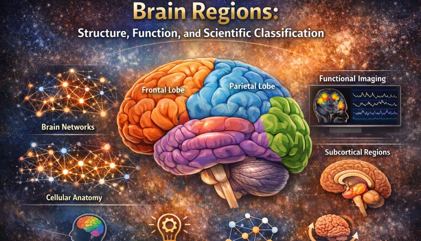
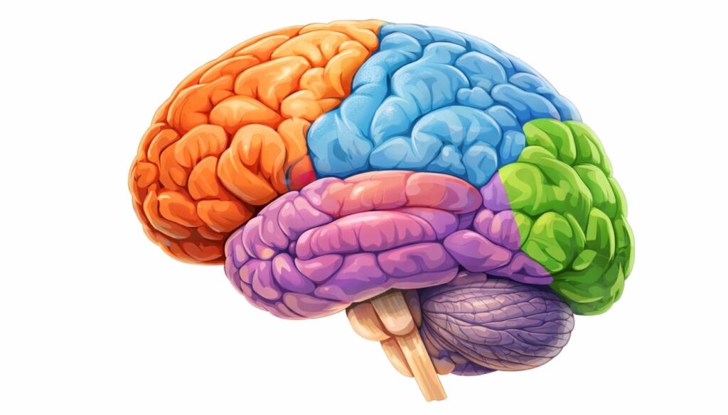
The cerebral cortex is divided into four primary lobes:

  • Frontal lobe – executive functions and voluntary movement

  • Parietal lobe – sensory integration and spatial processing

  • Temporal lobe – memory, language comprehension, and emotion

  • Occipital lobe – visual processing

Each lobe contains multiple specialized subregions.
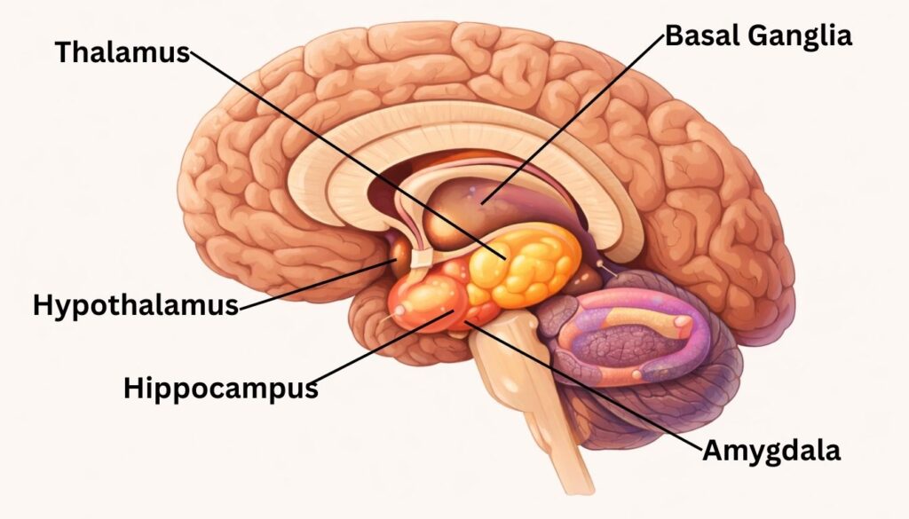
Subcortical regions lie beneath the cerebral cortex and include structures such as:

  • Thalamus

  • Hypothalamus

  • Basal ganglia

  • Amygdala

  • Hippocampus

These areas regulate motor control, memory, emotion, autonomic function, and sensory relay.

The thalamus receives nearly all sensory information (except smell) and relays it to the appropriate cortical areas. It also filters and modulates incoming signals, influencing attention and awareness. It acts as a gateway between sensory systems and conscious processing.

The prefrontal cortex supports planning, decision-making, impulse control, working memory, and social behavior. It integrates information from multiple brain regions to guide goal-directed behavior. It is one of the last brain areas to fully mature.

The hippocampus is essential for forming new declarative memories (facts and events). It consolidates short-term memories into long-term storage. Damage to this region can impair the ability to form new memories (anterograde amnesia).

Neuroplasticity refers to the brain’s ability to reorganize its structure and function in response to learning, experience, or injury. Brain regions can strengthen connections, form new synapses, and even reassign functions following damage.

Yes. Through functional reorganization, other regions may partially compensate for damaged areas. This process depends on age, extent of injury, and rehabilitation. However, compensation is often incomplete.

Some regions play central roles in emotional processing, such as the amygdala for threat detection and fear. However, emotions arise from distributed networks involving the limbic system, prefrontal cortex, and other regions. No emotion is controlled by a single isolated area.

No. While certain functions are lateralized (e.g., language often left-dominant), personality traits are not determined by hemisphere dominance. Both hemispheres work together for most cognitive processes.

Techniques like fMRI, PET, and SPECT allow researchers to observe activity and receptor distribution in living brains. Functional connectivity analysis can define regions based on synchronized activity rather than anatomical landmarks alone.

There is natural anatomical variability between individuals. Genetics, development, and environmental influences shape regional size and connectivity. This variability challenges rigid region-based models.

Research shows that cognition and behavior arise from coordinated activity across distributed systems. Network-based models better explain complex functions such as consciousness, attention, and self-referential thought than isolated regional models.

Conclusion

The study of brain regions provides a foundational framework for understanding the intricate workings of the human brain. From the broad anatomical divisions of the cortical lobes to the specific functional roles of subcortical structures, the concept of specialized brain areas has been instrumental in advancing our knowledge of neuroscience. However, as research progresses, it becomes increasingly clear that a purely region-based model is insufficient. The brain is not a collection of independent modules, but a highly integrated and dynamic network.
 
The shift towards a network-based perspective, which emphasizes the importance of functional connectivity and the coordinated activity of distributed brain regions, offers a more holistic and accurate model of brain function. This view acknowledges the complexity of the brain, where functions emerge from the dynamic interplay of multiple areas. 
 
It also accommodates the remarkable capacity of the brain for plasticity, allowing for both structural and functional reorganization in response to experience and injury. As we continue to refine our methods and technologies, our understanding of the brain’s regional and network architecture will undoubtedly evolve.
 
Ultimately, a comprehensive understanding of the brain requires a synthesis of both structural and network-based models. It is in the interplay between specialized regions and their dynamic connections that the richness of human cognition, emotion, and behavior emerges. Therefore, it is with a sense of scientific caution and an appreciation for the brain’s profound complexity that we should approach the study of its regions.
 
The journey to fully understanding the brain is far from over, and the concepts discussed here represent our current understanding in this ever-evolving field.
 

Disclaimer

 
This article is for educational and informational purposes only and does not constitute medical, psychological, or legal advice.
 

References


Share this post :

Facebook
Twitter
LinkedIn
Pinterest
Popular Product